
╔════════════════════════════════════════════════════════════════════╗
║ – Amniotic band syndrome is a rare pregnancy-related condition in which tissue bands can constrict developing fetal structures. ║
║ – The effects range from minor skin indentations to missing digits, limb differences, facial abnormalities or serious body-wall defects. ║
║ – Prenatal ultrasound can identify indirect signs, although the bands themselves are often difficult to see. ║
║ – Most cases are managed with monitoring during pregnancy and specialized treatment after birth, while fetal surgery is considered only in selected severe cases. ║
║ – The outlook depends on which structures are affected, how severe the constriction is and whether blood flow or vital organs are compromised. ║
╚════════════════════════════════════════════════════════════════════╝
What Is Amniotic Band Syndrome?
Amniotic band syndrome is a rare congenital condition that can develop when strands of tissue associated with the amniotic sac become attached to or wrap around parts of a developing fetus. The bands may restrict normal growth, movement or blood circulation, potentially producing abnormalities that vary from barely noticeable to life-threatening.
The condition is also known as amniotic band sequence, constriction band syndrome and amniotic band disruption complex. It is not a single predictable birth defect. Instead, doctors use the term to describe a group of physical abnormalities believed to result from fetal entanglement or disruption during development.
One of the most important characteristics of the condition is its wide range of severity. A baby may be born with nothing more than a narrow ring around a finger or toe, while another may have a shortened limb, missing digits, facial abnormalities or major defects involving the chest or abdomen.
The location and tightness of a band are particularly important. A loose band may leave an indentation without significantly affecting development. A tight band can compress blood vessels, nerves and other tissues and potentially cause permanent damage.
What Causes the Condition?
The most widely accepted explanation involves disruption of the amnion, the inner membrane surrounding the developing fetus. Following damage to this membrane, fibrous strands may form and become entangled with fetal structures.
As pregnancy progresses and the fetus grows, a strand can tighten around a developing finger, toe, arm, leg or another structure. If circulation becomes restricted, tissues beyond the constriction may not receive adequate oxygen and nutrients.
Researchers have also proposed other mechanisms for some abnormalities grouped under this condition. Vascular disruption and developmental disturbances may contribute to certain cases, which is why the term “sequence” is frequently used.
There is generally no evidence that ordinary activities during pregnancy cause the condition. Most cases occur sporadically rather than following a predictable inherited pattern.
Is It a Genetic Disorder?
In most cases, this condition is not regarded as a conventional inherited genetic disorder.
That distinction can be reassuring for families who may immediately wonder whether they passed something to their baby. The majority of cases are considered isolated events, and there is not usually a strong pattern of recurrence within families.
However, doctors may recommend additional evaluation when a fetus has several unusual abnormalities. Not every limb, facial or body-wall defect is caused by an amniotic band. Other congenital conditions can sometimes look similar on prenatal imaging.
A maternal-fetal medicine specialist can therefore evaluate the overall pattern of abnormalities rather than relying on one ultrasound finding.
Which Body Parts Can Be Affected?
The hands, fingers, feet and toes are common sites of involvement, but the condition is not limited to the extremities.
Possible findings include:
- Narrow constriction rings around fingers or toes
- Deep grooves around an arm or leg
- Missing portions of fingers, toes or limbs
- Shortened or underdeveloped extremities
- Webbing between digits
- Swelling caused by impaired circulation or lymphatic drainage
- Abnormal positioning of a limb or foot
- Cleft lip or palate
- Craniofacial abnormalities
- Chest-wall defects
- Abdominal-wall defects
- Abnormalities involving internal organs
- Rare involvement of the umbilical cord
The effects are determined by where a band attaches and how much pressure it places on developing tissues.
Symptoms and Signs
The physical signs depend on the severity and location of the restriction.
A superficial band may leave a circular indentation around a finger, toe or limb. The affected area may otherwise function normally.
A deeper constriction can interfere with circulation and produce swelling beyond the band. If blood flow remains restricted for a prolonged period, tissue development can be seriously affected.
Some babies may have a portion of a finger, toe or limb that did not develop normally. In other situations, the band may have caused tissue loss before birth, resulting in what appears to be a congenital amputation.
More complex cases may involve the face, skull, chest or abdomen. These abnormalities can have considerably greater medical consequences than an isolated limb constriction.
How Is the Condition Diagnosed During Pregnancy?
Prenatal ultrasound is the primary method used when doctors suspect the condition.
The actual fibrous bands can be extremely thin and difficult to visualize. As a result, physicians often identify the condition indirectly by looking for abnormalities in fetal anatomy, restricted movement, unusual positioning or swelling.
A detailed anatomy ultrasound can help determine:
- Which body parts are affected
- Whether a constriction ring is present
- Whether fetal movement is restricted
- Whether blood flow appears compromised
- Whether other structural abnormalities are present
- Whether the umbilical cord appears normal
In complex cases, fetal MRI may provide additional information. MRI is not required for every suspected case, but it can be useful when doctors need a clearer picture of complicated anatomy or are evaluating whether an intervention might be appropriate.
Importantly, seeing a membrane or strand on an ultrasound does not automatically establish the diagnosis. Specialists consider the entire clinical and imaging picture because other normal or abnormal membranes can sometimes resemble an amniotic band.
When Can It Be Detected?
Some abnormalities may become visible during early pregnancy, but detection is not guaranteed.
In many pregnancies, concerns become more apparent during the detailed anatomy examination performed around the middle of pregnancy. A clinician may notice an abnormal limb position, swelling, a missing digit or another structural difference.
Some cases are not recognized until delivery because the band itself is too small to see clearly on prenatal imaging.
This means a normal early ultrasound does not necessarily rule out every future structural abnormality.
What Happens After a Prenatal Diagnosis?
A suspected diagnosis usually leads to more detailed evaluation rather than an immediate treatment decision.
Maternal-fetal medicine specialists may perform additional ultrasounds and monitor the baby’s growth, movement and affected structures. The medical team may also assess whether the condition appears stable or whether there are signs of progressive constriction.
The delivery plan depends on the abnormalities involved.
A baby with an isolated limb difference may not require a highly specialized delivery environment. A fetus with significant chest-wall, abdominal or other organ abnormalities may benefit from delivery at a center with neonatal intensive care and pediatric surgical expertise.
Parents may also meet specialists before delivery so that treatment can be organized in advance.
Can It Be Treated Before Birth?
Most cases are not treated surgically during pregnancy.
The primary approach is individualized monitoring and planning for treatment after delivery. However, fetal intervention can be considered in rare situations where a constricting band threatens an important fetal structure.
Fetoscopic band release is a minimally invasive fetal procedure in which physicians enter the uterus with specialized instruments and attempt to release the constricting tissue.
The goal is to prevent additional injury, preserve blood flow and, in selected cases, protect an endangered limb.
Fetal surgery is not appropriate simply because an amniotic band is visible. The potential benefit must outweigh significant risks to both the pregnant patient and fetus.
Recent evidence remains limited because the condition is uncommon and reported fetal-surgery cases are relatively small. A systematic review of published fetoscopic-release cases found potential benefits for preserving affected limbs, but also reported substantial risks, including premature rupture of membranes and preterm birth.
For that reason, decisions about fetal intervention are made by highly specialized fetal therapy teams after detailed imaging and multidisciplinary assessment.
What Treatment Is Available After Birth?
Postnatal treatment depends on the specific abnormality.
Some babies require no treatment. A shallow constriction ring that does not interfere with circulation, movement or function may simply be observed.
Other children may need surgery.
Reconstructive Surgery
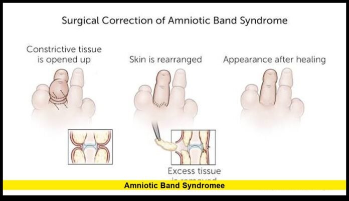
Surgical procedures can release constriction rings and improve the appearance or function of affected tissues.
Depending on the abnormality, reconstructive treatment may address:
- Tight tissue bands
- Webbed fingers or toes
- Limb deformities
- Clubfoot
- Cleft lip or palate
- Chest-wall abnormalities
- Abdominal-wall defects
- Other structural differences
Timing varies. Some procedures are performed during infancy, while others are delayed until the child is older.
Prosthetics
Children who are born with missing or severely shortened limbs may benefit from prosthetic devices.
Modern pediatric prosthetics can be adjusted as children grow. In some situations, prosthetic treatment is introduced when the child is developmentally ready to use the device effectively.
The goal is not simply cosmetic. Prosthetic care can support mobility, balance, grasping and independence depending on the affected limb.
Physical and Occupational Therapy
Rehabilitation can be an important part of long-term care.
Physical therapy may help improve strength, balance, mobility and coordination. Occupational therapy can focus on daily activities, hand function and adapting tasks to a child’s individual abilities.
Children often learn effective ways to perform everyday activities even when they have significant limb differences.
Is Fetal Surgery Always Successful?
No. Fetal surgery cannot reverse damage that has already occurred.
If a band has severely damaged tissue or interrupted circulation for a prolonged period, releasing it may not restore normal anatomy.
The potential benefit is greatest when doctors believe the band is actively threatening a structure that could still be preserved.
Fetoscopic treatment also carries significant pregnancy risks. Premature rupture of membranes is one of the major concerns and can lead to premature delivery.
Therefore, fetal surgery is generally reserved for carefully selected cases rather than used as routine treatment.
Can Babies Survive the Condition?
Yes. Many babies with this condition survive, particularly when abnormalities are limited to fingers, toes or a single limb.
The prognosis becomes more serious when the condition affects vital organs or the umbilical cord.
An isolated constriction ring can have little impact on life expectancy. By contrast, extensive fetal injury, major body-wall defects or severe umbilical cord involvement can create life-threatening complications.
Doctors therefore avoid giving families a prognosis based solely on the diagnosis. Instead, they examine the specific structures involved and the degree of damage.
What Is the Long-Term Outlook?
The long-term outlook varies considerably.
Children with mild abnormalities may require little medical intervention and can have normal life expectancy and good function.
Children with limb differences may need reconstructive surgery, prosthetic devices, therapy or a combination of these approaches. Many adapt extremely well as they grow.
More complicated abnormalities may require multiple surgeries and long-term multidisciplinary care.
The child’s treatment plan can also evolve over time. A procedure that is useful during infancy may be followed by another intervention during childhood as bones, muscles and soft tissues develop.
Can It Be Prevented?
There is currently no established way to prevent most cases.
Because the underlying mechanism is not completely understood and many cases occur sporadically, there is generally no specific pregnancy behavior that can be changed to eliminate the risk.
Routine prenatal care remains important because ultrasound examinations can identify many structural abnormalities and allow families to receive specialist counseling when concerns arise.
Questions Parents May Want to Ask
Receiving an unexpected prenatal diagnosis can be overwhelming. Parents may want to ask their healthcare team:
- Which parts of the baby are affected?
- Is blood flow to the affected area normal?
- Is the condition expected to progress?
- Are any internal organs involved?
- Does the baby need additional imaging?
- Is fetal intervention an option?
- What are the risks of fetal surgery?
- Where should delivery take place?
- Which specialists will be involved after birth?
- Will surgery, prosthetics or rehabilitation be necessary?
- What is the expected long-term functional outlook?
Getting answers to these questions can help families understand what the diagnosis means for their particular pregnancy rather than relying on general descriptions.
Why Specialist Evaluation Is Important
The condition can look very different from one pregnancy to another. A small indentation on a digit and a complex body-wall abnormality may fall under the same broad diagnostic category while requiring completely different treatment.
Maternal-fetal medicine specialists can evaluate prenatal findings, while fetal therapy teams can determine whether an in-utero procedure might provide meaningful benefit.
After birth, pediatric surgeons, orthopedic specialists, plastic surgeons, rehabilitation professionals, prosthetists and other clinicians may become involved depending on the baby’s needs.
The most important consideration is creating a treatment strategy around the child’s actual anatomy and functional needs.
The Bottom Line
Amniotic band syndrome is an uncommon congenital condition with an exceptionally broad range of possible effects. It can produce minor constriction marks or significant developmental abnormalities involving the limbs, face, body wall, internal organs or umbilical cord.
Prenatal diagnosis can be difficult because the bands themselves are often too small to see. Instead, doctors frequently recognize the condition through its effects on fetal anatomy, movement or blood flow.
For most pregnancies, careful monitoring and preparation for treatment after birth are the main approaches. In carefully selected severe cases, fetoscopic release may be considered when doctors believe a developing limb or other structure remains at risk.
Ultimately, the diagnosis alone does not determine a child’s future. The location and severity of the abnormality, the structures involved and the availability of specialized treatment all influence the outcome. With modern pediatric surgery, prosthetics and rehabilitation, many children with isolated abnormalities can develop strong functional independence and lead active lives.
Have questions or experiences about this rare pregnancy condition? Join the conversation below and stay tuned for more trustworthy health and pregnancy updates.